
Figure 2. a, b.
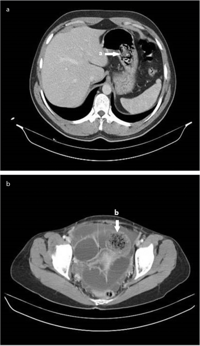
CT scan showing a phytobezoar. Typical mottled appearance of a phytobezoar in the stomach (a) and distal ileal bowel loops (b)
Official websites use .gov
A
.gov website belongs to an official
government organization in the United States.
Secure .gov websites use HTTPS
A lock (
) or https:// means you've safely
connected to the .gov website. Share sensitive
information only on official, secure websites.
CT scan showing a phytobezoar. Typical mottled appearance of a phytobezoar in the stomach (a) and distal ileal bowel loops (b)