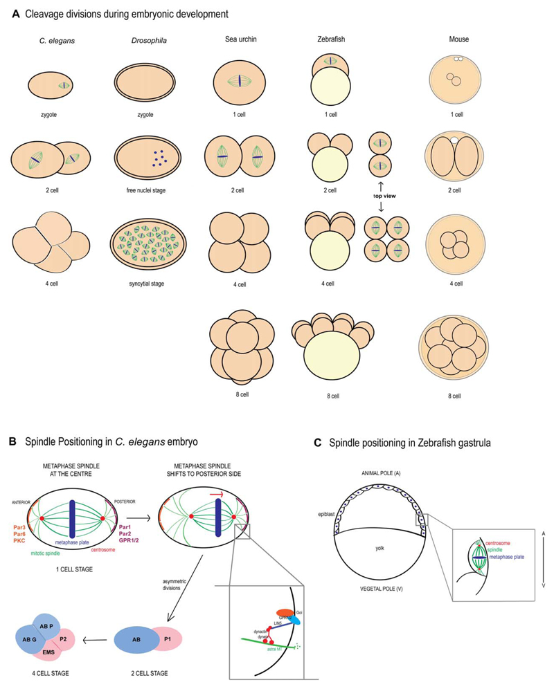
Fig 2. Cleavage divisions across metazoa.
A: Representation of spindle positioning from zygote (1 cell) to 8 cell stage in various metazoans. In the one-cell stage C. elegans embryo, the spindle is positioned asymmetrically toward the posterior end, giving rise to daughter cells with different fates. In Drosophila, successive nuclear divisions coupled with the absence of cytokinesis give rise to a syncytial embryo. Sea urchin embryos undergo asymmetric divisions giving rise to micromeres and macromeres. In contrast, early divisions are symmetric in zebrafish embryos. Mouse embryos also undergo asymmetric divisions, giving rise to daughter cells with different cell fates. B: In the one-cell stage C. elegans embryo, the mitotic spindle shifts to the posterior end, giving rise to AB and P1 cells, which again undergo asymmetric divisions. C: During gastrulation in zebrafish, spindles are positioned along the animal-vegetal axis.

