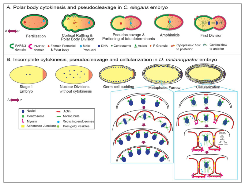
Fig 4. Variations in cytokinesis during metazoan development.
A: Fertilization-triggered meiotic divisions of the oocyte in C. elegans are extremely asymmetric resulting in polar bodies devoid of cytoplasm and the haploid oocyte retaining most of the cytoplasm. Soon after fertilization, cortical contraction in the C. elegans embryo forms a pseudocleavage that segregates the fate determinants in the zygote, a process that is essential for antero-posterior axis specification. B: Drosophila embryos initially demonstrate a cytokinesis-free nuclear division upto 8 nuclear cycles. At the 9th nuclear division, these nuclei migrate to the periphery and continue to divide without undergoing cytoplasmic divisions for another three cycles. During these peripheral divisions, they form pseudocleavages/partial furrows to shield the chromatin from the influence of neighboring asters. The nuclei finally undergo cellularization at interphase of the 14th nuclear division, utilizing an actomyosin-based contractile machinery similar to conventional cytokinesis.

