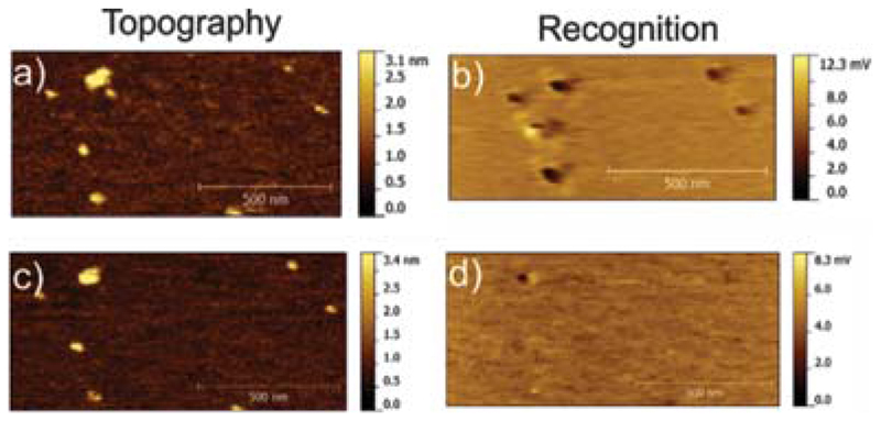
Fig. 5.
Block experiment. (a and b) Topography and recognition images of UCP1 molecules in the lipid membrane acquired with an ATP-tethered tip, prior to blocking. (c and d) Topography and recognition images after blocking by addition of free ATP into the solution while scanning the same position. Almost all recognition spots (black) disappeared, demonstrating the specificity of the UCP1-ATP interaction.

