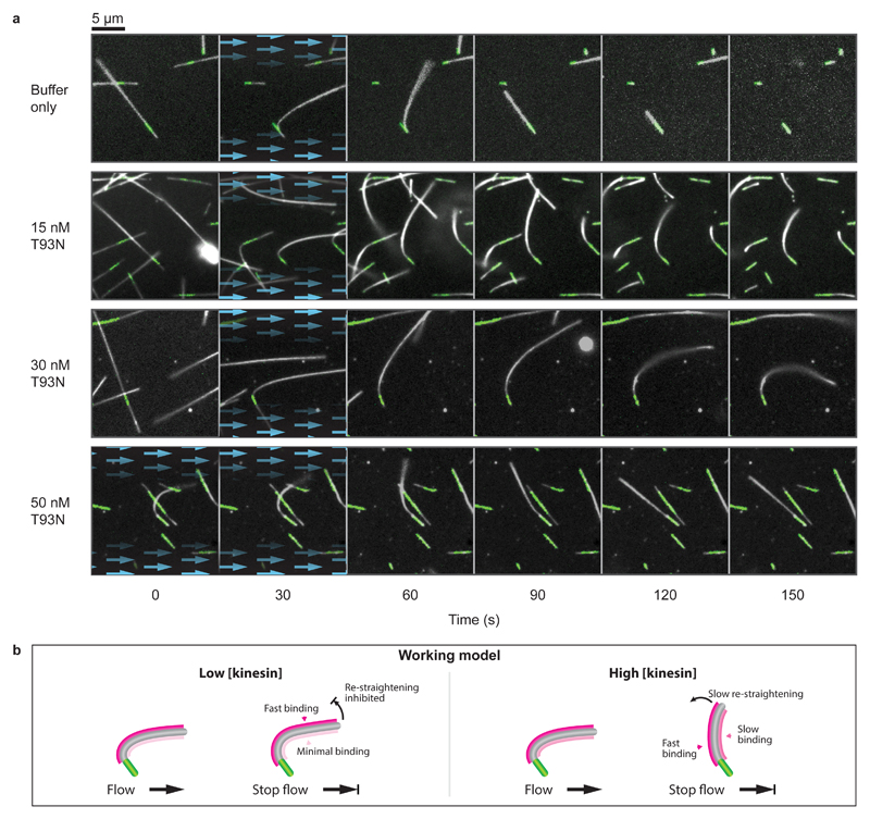
Figure 4. Nucleotide-free motor domains can bend-lock microtubules.
a, Time-lapse images of microtubule bending experiments for a range of kinesin concentrations. Blue arrows highlight the presence and direction of fluid flow. Dynamic microtubules appear white (dark-field) and fluorescent seeds are marked in green (epi-fluorescence). Each condition was tested twice on independent occasions. Microtubules shown here have been selected for having similar orientations. A more extensive selection is given in Supplementary Movie 2, which shows a complete range of orientations and lengths. b, Working model. We propose that kinesin binds preferentially to the stretched (convex) side of the microtubule and also stabilises this expanded region of the microtubule lattice. At high kinesin concentrations, the convex side of the microtubule would quickly saturate. Binding would also occur slowly on the concave side, causing this side to expand, progressively re-straightening the microtubule.

