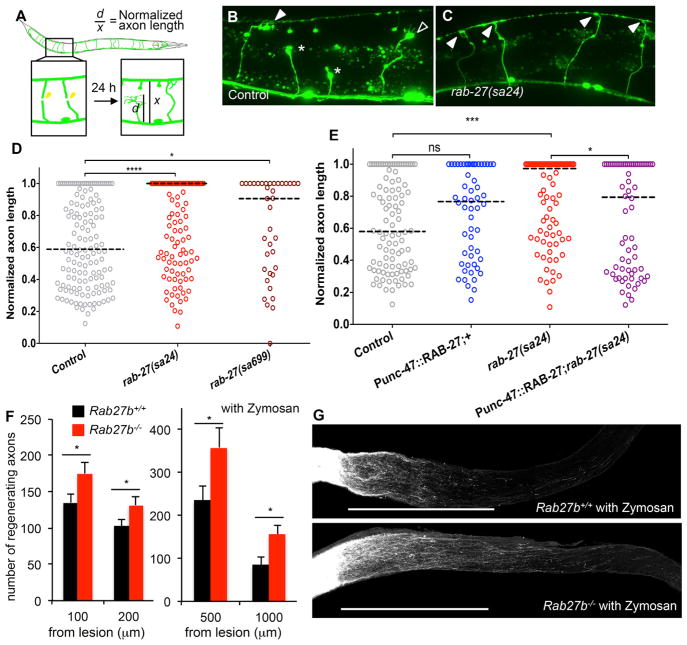
Figure 6. Rab27 Inhibits Axonal Regeneration In Vivo.
(A) Commissural axons of the GABAergic DD/VD neurons are severed using a pulsed laser, and regeneration is assessed after 24 hr in young adult (L4 stage + 24 hr at 20°C) animals.
(B) Normalized axon length in control and rab-27 mutant animals. Number of axons cut per genotype, left to right: 142, 148, and 37.
(C and D) Regenerating GABA axons 24 hr after axotomy in control (C) and rab-27-null (D) animals. Filled arrows indicate fully regenerated axons reaching the dorsal nerve cord, empty arrows indicate partial regeneration, and stars indicate nonregenerating axon stumps. All animals express Punc-47::GFP, which drives GFP expressing specifically in the GABA motor neurons.
(E) Normalized axon length in control, rab-27 mutants, and animals specifically expressing rab-27 cDNA in GABA neurons, in control and rab-27 mutant animals. Number of axons cut per genotype, left to right: 98, 56, 84, and 68. Kolmogorov-Smirnov test was used. ns, not significant; *p < 0.05, ***p < 0.0005, ****p < 0.0001.
(F) Age-matched (9–10 weeks old without zymosan, 14 weeks old with zymosan) animals underwent optic nerve crush (ONC). Quantification of regenerating RGC axons at indicated distances distal to the lesion sites at 17 days after injury from WT control mouse and Rab27b −/− mice. Data are presented as mean with SEM. Without zymosan, n = 29 Rab27b+/+ and n = 28 Rab27b−/−, and with zymosan, n = 10 Rab27b+/+ and n = 8 Rab27b−/− mice. *p < 0.05, Student’s t test.
(G) Representative confocal images of optic nerve at 17 days after crush injury with zymosan injection from WT control mouse and Rab27b−/− mouse. The CTB-labeled RGC axons are white. The eye is to the left and the brain is to the right. Scale bars represent 500 μm.

