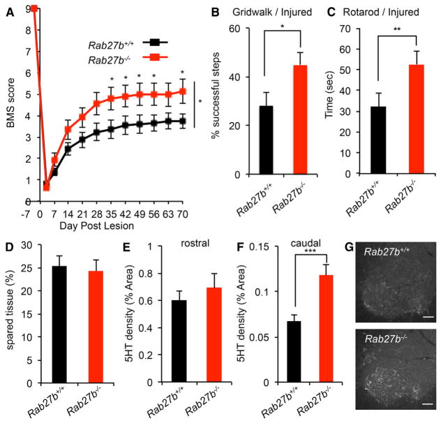
Figure 7. Improvement of Functional Recovery after Spinal Cord Injury in Rab27b−/− Mouse.
(A) Open-field locomotion performance measured by BMS of Rab27b+/+ and Rab27b−/− mice. Animals were scored on day post-lesion (DPL) −3, 3, 7, 14, 21, 28, 35, 42, 49, 56, 63, and 70 by two experienced observers blinded to group. Data are mean ± SEM for n = 19 Rab27b+/+ and n = 17 Rab27b−/−. *p < 0.05, significant difference between genotypes through DPL 35 to 70, one-way repeated-measure ANOVA across time series followed by Student’s t test between genotypes at indicated times.
(B and C) Gridwalk test at DPL 55 (B) and RotaRod performance at DPL 48 (C) of Rab27b+/+ and Rab27b−/− mice. Data are mean with SEM for n = 19 Rab27b+/+ and n = 17 Rab27b−/−. *p < 0.05 and **p < 0.01, Student’s t test.
(D) Sagittal sections of thoracic cord were stained with anti-GFAP antibody, and the extent of spared tissue at the injury site was quantified. Data are presented as mean with SEM for n = 19 Rab27b+/+ and n = 17 Rab27b−/−. No significant differences between groups with Student’s t test.
(E and F) Serotonergic (5HT+) fiber density at coronal sections of rostral to the lesion (E) and caudal to the lesion (F) from Rab27b+/+ and Rab27b−/− mice 70 days after hemisection were quantified. Data are presented as mean with SEM for n = 19 Rab27b+/+ and n = 17 Rab27b−/−. No significant differences between groups with Student’s t test (E). *p < 0.05, Student’s t test (F).
(G) Representative image of raphespinal fibers stained with anti-5HT antibody in the spinal ventral horn.

