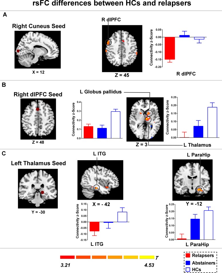
Fig 3. Group differences of rsFC existed between the seeds and the cluster location.
(A) Compared with HCs, Relapsers showed significantly increased negative functional connectivity between the right cuneus and the right dlPFC; (B) Compared with HCs, Relapsers showed decreased rsFC between the right dlPFC and the left thalamus, and the left globus pallidus; (C) Compared with HCs, Relapsers showed decreased rsFC between the left thalamus and the left inferior temporal gyrus, and the parahippocampal gyrus. Brain maps of representative slices of ROIs are also showed in the figure and colored dots represent seed locations. Bar graphs display mean rsFC z scores for abstainers, relapsers and HCs and the error bars represent standard deviation. Abbreviations: dlPFC, dorsolateral prefrontal cortex; ITG, inferior temporal gyrus; ParaHip, parahippocampal gyrus; HCs, healthy controls; rsFC, resting-state functional connectivity.

