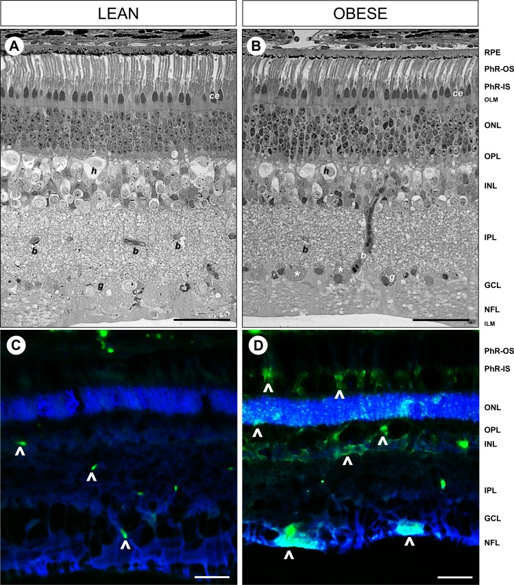
Figure 2.
Cellular derangement of retinal structure and degenerating neurons in the Western diet-fed Ossabaw pigs. Light microscopy of toluidine blue staining on 2-μm thick resin-embedded retinal sections revealed intact and organized cellular structures in (A) lean control pigs, which were disrupted in the (B) obese Ossabaw pigs. FJC staining to visualize degenerating neurons (arrowheads) in (C) lean and (D) obese Ossabaw pigs showed increased labeling of cell bodies in the nerve fiber layer (NFL), INL, and PhR of the obese animals. PhR-IS, photoreceptor inner segment; OLM, outer limiting membrane; ONL, outer nuclear layer; OPL, outer plexiform layer; ILM, inner-limiting membrane; g, ganglion cell; b, blood vessel; h, horizontal cell; ce, cone ellipsoid. n = 3 eyes per group. Scale bar: 50 μm.

