Figure 6.
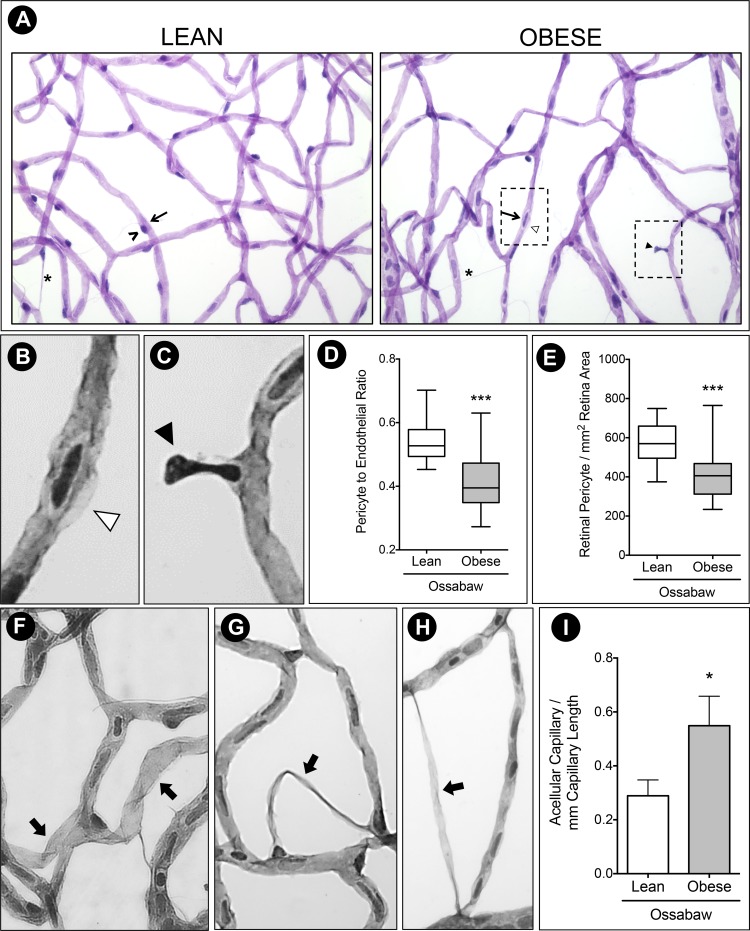
Pericyte ghost and acellular capillaries in Western diet-fed Ossabaw pigs. (A) Trypsin digest of Ossabaw pig retinas revealed uneven capillary caliber in the obese group, with pericyte lost (black arrowhead) and pericyte ghost (white arrowhead). Migrating pericytes (asterisk) can be seen in both animal groups. (B) Inset of pericyte ghost. (C) Inset of pericyte dropout. (D) Pericyte to endothelial ratio was significantly reduced in obese pigs compared to the lean control, evident by (E) decreased total retinal pericyte count in obese Ossabaw pigs. (F–H) Representative images of acellular capillaries (thick arrow) identified and quantified in Ossabaw pig retinas. (I) Count of acellular capillaries showed 50% increase per millimeter capillary length in Western diet-fed pigs. ^, pericyte cell; arrow, endothelial cell. n = 3 eyes per group. *P < 0.05 and ***P < 0.001.

