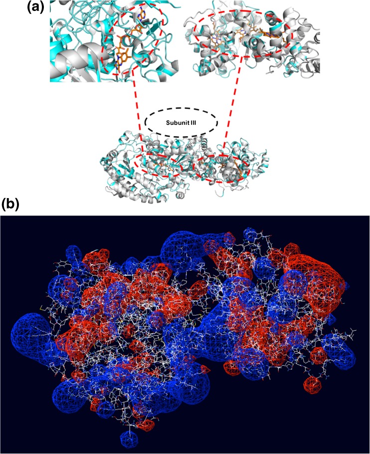
Fig. 5.
(a) Detailed representation of structural alignment between Subunit I of FDH (homology model, grey structure) and Aspergillus niger FAD-glucose dehydrogenase (PDB ID: 4ynt, light blue structure); and between Subunit II of FDH (homology model) and thiosulfate dehydrogenase (tsdba) from Marichromatium purpuratum “as2 isolated” form (PDB ID: 5lo9); (b) electrostatic map of Subunits I and II after docking obtained from the homology models

