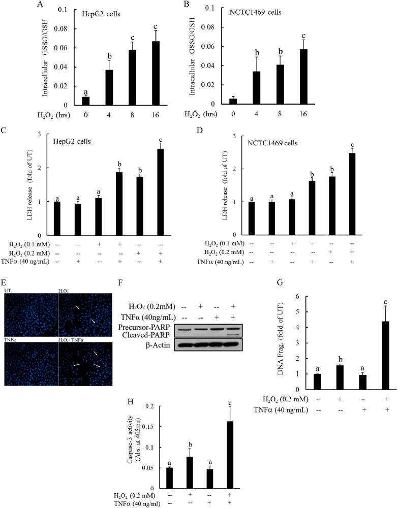
Fig. 2. Intracellular glutathione imbalance sensitizes hepatocytes to TNFα cytotoxicity.
a, b Both HepG2 (a) and NCTC1469 cells (b) were exposed to complete DMEM containing H2O2 (0.2 mM) for the indicated time periods. Intracellular GSH and GSSG levels were measured, and the GSSG/GSH ratios were calculated. All values are denoted as the mean ± SD from three or more independent studies. Bars with different characters differ significantly (p < 0.05). c, d H2O2 sensitizes hepatocytes to TNFα-induced cell death. HepG2 (c) and NCTC cells (d) were pretreated with H2O2 (0.1 and 0.2 mM) for 2 h before the addition of TNFα (40 ng/mL). Cell death was measured 16 h later by LDH release assay. Bars with different characters differ significantly (p < 0.05). e Hoechst 33342 staining. Arrows denote apoptotic bodies. f Western blot analysis of PARP cleavage. HepG2 cells were pretreated with H2O2 (0.2 mM) for 2 h, followed by TNFα (40 ng/mL) stimulation. Whole-cell lysates were collected and subjected to western blot for the detection of PARP cleavage. g DNA fragmentation ELISA assay. h Caspase-3 activities. Bars with different characters differ significantly (p < 0.05)

