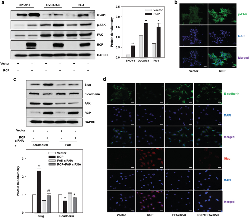
Fig. 1. FAK is important for RCP-induced ovarian cancer cell EMT.
a Ovarian cancer cells were transfected with the indicated vectors for 48 h. Immunoblotting (left). Immunoblot bands were quantified by Image J densitometric analysis and normalized to the control vector (right; mean ± s.d. *P < 0.05, **P < 0.01 vs. the control vector). b PA-1 cells were transfected with the indicated vectors, and the expression of FAK phosphorylation was visualized by immunofluorescence. Original magnification, ×200; scale bar, 20 μm. Representative results are presented from at least three independent experiments with similar results. c SKOV-3 cells were co-transfected with the indicated vectors and siRNAs for 48 h. Immunoblotting (upper). Densitometric analysis (lower; mean ± s.d. **P < 0.01 vs. the control vector, #P < 0.05, ##P < 0.01 vs. RCP overexpression with scrambled siRNA). d SKOV-3 cells were transfected with the indicated vectors for 48 h, serum-starved, and treated with PF573228 (10 μM) for 1 h, and the expression of E-cadherin and Slug was visualized by immunofluorescence. Original magnification, ×200; scale bar, 20 μm. All experiments were repeated three times

