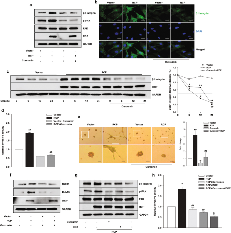
Fig. 5. Curcumin inhibits stabilization of β1 integrin protein and FAK phosphorylation.
a SKOV-3 cells were transfected the indicated vectors for 48 h, serum-starved, and treated with curcumin (20 μM) for 2 h. Immunoblotting. b The expression of β1 integrin was visualized by immunofluorescence. Original magnification, ×400; scale bar, 20 μm. c SKOV-3 cells were transfected with the indicated vectors for 48 h, serum-starved, and pretreated with curcumin (20 μM) for 2 h and then incubated with CHX (20 μg/ml) for the indicated times. Immunoblotting (left). Densitometric analysis (right; mean ± s.d. *P < 0.05, **P < 0.01 vs. the control vector, #P < 0.05, ##P < 0.01 vs. RCP overexpression only). d SKOV-3 cells were transfected with the indicated vectors for 48 h followed by treatment with curcumin (20 μM) for 1 h. Invasion assay (mean ± s.d. **P < 0.01 vs. the control vector, ##P < 0.01 vs. RCP overexpression only). e Phase contrast images showing the morphology of stably transfected SKOV-3 cells with the indicated vectors followed by treatment with curcumin (20 μM) for 4 days on Matrigel. Original magnification, ×100; scale bar, 200 μm. Inset shows higher magnification (left). The number of branches was counted and quantified (right); mean ± s.d. **P < 0.01 vs. the control vector, ##P < 0.01 vs. RCP overexpression only. f SKOV-3 cells were transfected with the indicated vectors for 48 h, serum-starved, and treated with curcumin (20 μM) for 1 h. Immunoblotting. g SKOV-3 cells were transfected with the indicated vectors for 48 h, serum-starved, and treated with curcumin (20 μM), DOX (0.001 μM), and curcumin with DOX. Immunoblotting and h invasion assay (mean ± s.d. *P < 0.05 vs. the control vector, ##P < 0.01 vs. RCP overexpression only, $P < 0.05 vs. RCP overexpression and DOX treatment). All experiments were repeated three times

