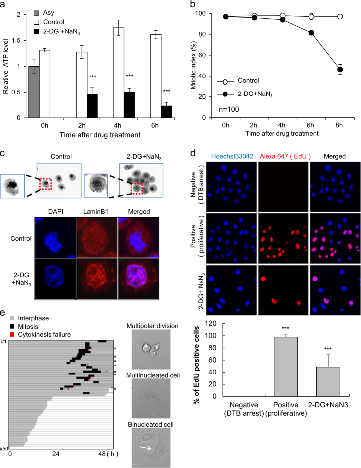
Fig. 1. Induction of mitotic slippage by ATP depletion.
Cells incubated with 100 ng/ml nocodazole for 16 h were treated with/without 6 mM 2-deoxyglucose (2-DG) and 10 mM sodium azide (NaN3). a The relative ATP levels of cells were measured at the indicated time points after 2-DG and NaN3 treatment. The results are given as the mean ± SD from three independent experiments. ***P < 0.001 by Student’s t-test. b Quantification of the percentage of mitotic cells with condensed chromosome was performed by aceto-orcein staining. The results are given as the mean ± SD from three independent experiments (n = 100). c Images of cells with condensed or decondensed chromosomes 8 h after 2-DG and NaN3 treatment by aceto-orcein staining (upper panel). Immunocytochemistry images of reassembled nuclear membrane were obtained by lamin B staining with DAPI (lower panel). d The EdU incorporation assay was performed using cells that underwent mitotic slippage after 8 h of treatment with 2-DG and NaN3. After washing twice with PBS, attached cells were cultured with an EdU solution for 16 h in fresh medium with nocodazole. EdU-positive cells were detected by the red fluorescence signal (upper panel). Quantification of the percent of EdU-positive cells (lower panel). The results are given as the mean ± SD from three independent experiments. ***P < 0.001 by Student’s t-test. e The conditions used were the same as described for the EdU incorporation assay. Monitoring the fate of attached cells by ATP depletion was performed in the absence of nocodazole for 48 h (left panel). *multipolar division. Representative images of multipolar division, multi-nucleated cells, and binucleated cells (white arrow) (right panel). Scale bar, 10 µm

