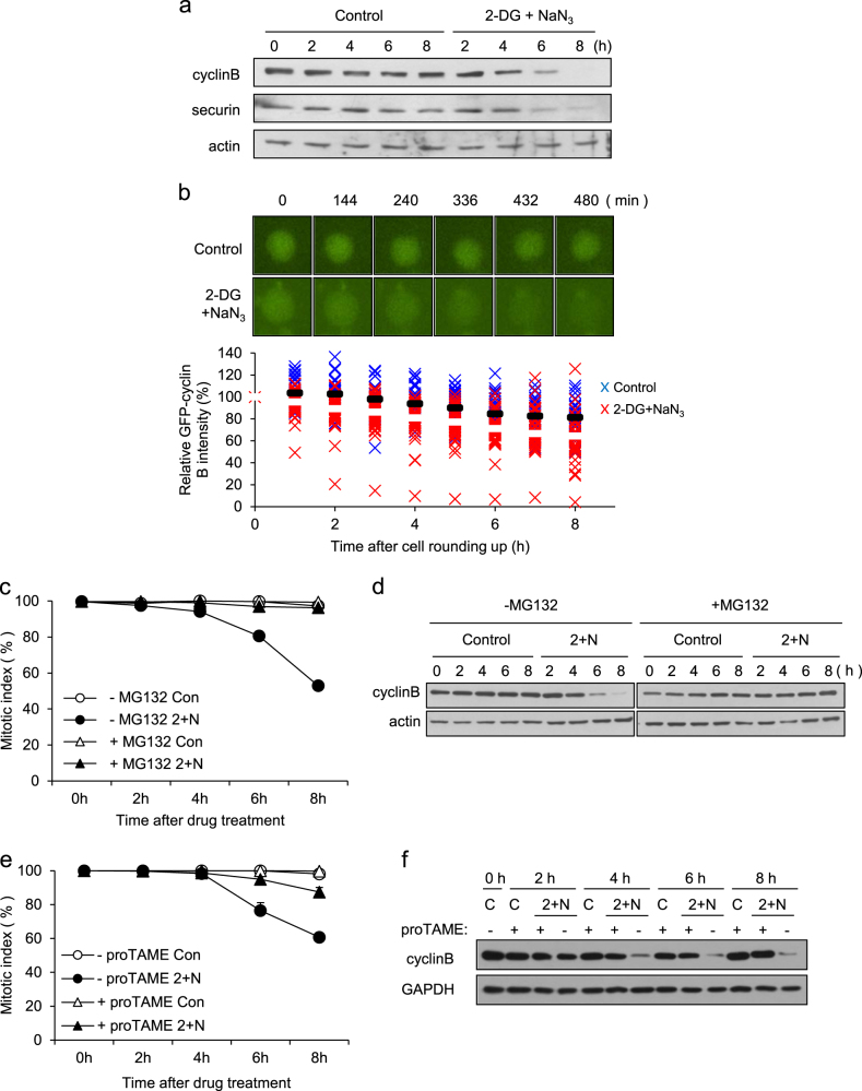
Fig. 2. Mitotic slippage via APC/C-dependent degradation of cyclin B.
a Mitotic arrested cell lysates from the indicated times after treatment with/without 2-DG and NaN3 were subjected to western blot analysis using antibodies for cyclin B, securin, and actin (loading control). b Cell arrested in mitosis were transfected with GFP-cyclin B and treated with/without 2-DG and NaN3 in the presence of nocodazole. Time-lapse images of cyclin B degradation are representative of 36 control cells and 47 cells treated with 2-DG and NaN3. Time is presented as minutes after cell rounding up for mitosis. The relative intensity of the GFP-cyclin B signal at different times was measured using NIS-Elements Viewer 4.0 software. The results are shown as the mean (control; black bar, 2-DG and NaN3; white bar). c Cells arrested in mitosis were treated with/without 2-DG and NaN3 (2 + N) in the absence/presence of MG132. Quantification of the percentage of mitotic cells with condensed chromosomes was performed by aceto-orcein staining. The results are shown as the mean ± SD from two independent experiments (n = 100). d Cell lysates from the indicated times after treatment with/without 2-DG and NaN3 (2 + N) were subjected to western blot analysis using antibodies against cyclin B and actin (loading control). e For APC inhibition, cells arrested in mitosis were treated with 12 μM proTAME for 30 min prior to co-treatment with/without 2-DG and NaN3 (2 + N). Quantification of the percentage of mitotic cells with condensed chromosome was performed by aceto-orcein staining. The results are shown as the mean ± SD from two independent experiments (n = 100). f Cell lysates from the indicated times after treatment with 2-DG and NaN3 (2 + N) were subjected to western blot analysis using antibodies for cyclin B and GAPDH (loading control)

