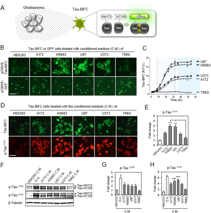
Fig. 2. Glioblastoma activates tau hyper-phosphorylation and aggregation.
a Illustration of glioblastoma-secretome effect on tau-BiFC sensor cells. As a fluorescence turn-on sensor, Venus fluorescence turns on only when tau assembles together. b The effect of glioblastoma conditioned media (C.M.) on tau aggregation. Tau-BiFC cells were treated with glioblastoma (A172, HS683, U87, U373, or T98G) conditioned media or HEK293-conditioned medium for 48 h. Stable cells expressing GFP were tested as a negative control, indicating that glioblastoma conditioned media did not activate CMV-promoter mediated gene expression. Scale bar, 100 µm. c Quantification of tau-BiFC fluorescence intensity. Error bars represent the standard deviations of three independent experiments. d, e Tau immuno-fluorescence images with p-Tau (Ser199) antibody and the quantification. The significance of the experiments was determined with Student’s t-test. ***p < 0.001. f Immuno-blot analysis of tau phosphorylation with p-Tau (Ser396) or anti-p-Tau (Ser199). For the immuno-blot analysis, tau-BiFC cells were treated with glioblastoma-C.M for 24 h. HEK293-conditioned medium was used as a negative control and okadaic acid (O.A.) was used as a positive control. g, h Quantification of p-Tau (Ser396) and p-Tau (Ser199). Error bars represent the standard deviations of three independent experiments. The significance of the experiments was determined with Student’s t-test. ***p < 0.001

