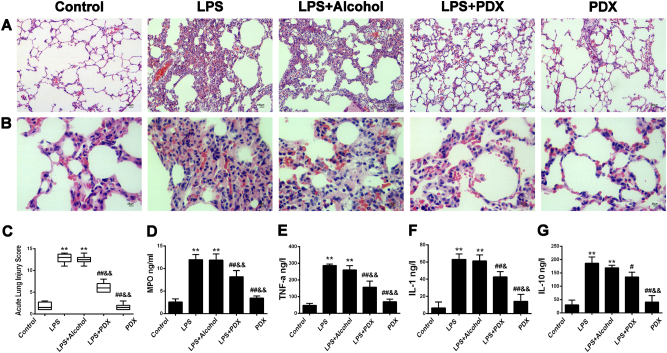
Fig. 1. Protectin DX protected lung tissues in LPS-induced acute lung injury.
Protectin DX (5 µg/kg) was injected into the caudal veins of Sprague-Dawley rats 8 h after LPS (14 mg/kg) stimulation; the rats were then ventilated for 60 min, and the effects of protectin DX were assessed (a, b) in hematoxylin and eosin-stained sections (original magnification ×100, ×400). Lung injury scores (c) were recorded from 0 (no damage) to 16 (maximum damage) according to the criteria described in the “Materials and methods”. Myeloperoxidase (d), TNF-α (e), IL-1 (f), and IL-10 (g) expression levels in lung tissues were measured by ELISA to quantitatively determine the resolution of infiltrated cells. The data are presented as the mean ± SD. n = 8. PDX protectin DX, MPO myeloperoxidase. Alcohol was the solvent for protectin DX. **p < 0.01 versus the control group; †p < 0.05, ††p < 0.01 versus the LPS group; ‡p < 0.05, ‡‡p < 0.01 versus the LPS + alcohol group

