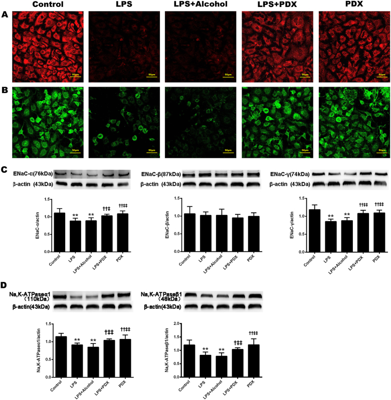
Fig. 6. Protectin DX promoted sodium channel expression in primary rat ATII cells stimulated with LPS.
Rat primary ATII cells were treated with protectin DX (3.605 × 10⁻³ mg/l) in the presence or absence of LPS (1 µg/ml) for 6 h. After incubation, the cells were harvested and sonicated. Sodium channel α (a) and Na, K-ATPase α1 (b) subunit protein expression levels in the cell lysates were detected by confocal laser-scanning microscopy using a specific Abs (original magnification ×400). In addition, sodium channel α, β, and γ subunit (c) and Na, K-ATPase α1 and β1 subunit (d) protein expression levels in the cell lysates were detected by western blotting. The data are presented as the mean ± SD. n = 8. PDX protectin DX. Alcohol was the solvent for protectin DX. **p < 0.01 versus the control group; †p < 0.05, ††p < 0.01 versus the LPS group; ‡p < 0.05, ‡‡p < 0.01 versus the LPS + alcohol group

