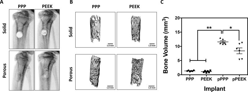
Figure 4. Bone growth throughout porous materials, especially PPP showed significantly increased bone ingrowth.
A) In vivo x-ray radiographs at 8-weeks post-surgery. (B) representative micro-CT reconstructions of same specimens from x-ray images. (C) Quantitative measurement of mineralized bone volume of implants. Solid scaffolds showed no significant difference of BV between groups. Porous PPP explants showed significantly increased BV value compared to any other explants. *p<0.05; **p<0.0001; n=6; data presented as mean ± S.E.M.

