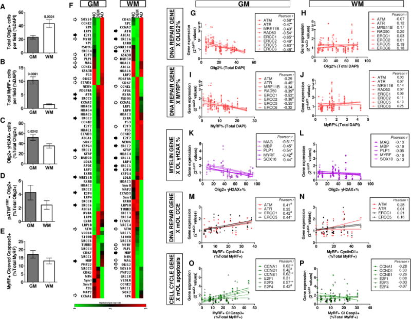
Fig. 4. Intracortical OL are more vulnerable to DNA damage and ectopic cell cycle associated degeneration than OL in the WM.

The regional differences in OL population between the intracortical region (GM) and subcortical WM were compared (n=24). A, B The OL (Olig2+) population was significantly higher in the subcortical WM, in contrast, the myelinating mOL (MyRF+) population was significantly higher in the GM. C The intracortical OL population suffer from a significantly higher degree of DSB and oxidative DNA damage. D, E Higher, but not significant, ATM activity and apoptosis were detectable the intracortical OL population in the GM (All unpaired t-test). F Gene expression profile clustergram of the intracortical GM and subcortical WM normalized against clinical phenotype (NC, DEM and AD) were compared. Open arrows: consistent trend of gene expression found between GM and WM in all groups. Closed arrows, opposing trend of gene expression found between GM and WM in all groups. Without arrows, genes with no consistent trend between GM and WM. G–P The differential histological data in GM and WM was plotted against the regional gene expression level for each subject. G–J The number of OL lineage and mOL population were negatively correlated with DNA repair gene expression. K–L The percentage of OL with DSB-DNA damage was negatively correlated with multiple myelin gene expression. M–N The percentage of mOL with CCE was directly proportional to DNA repair gene expression. O–P The expression of cell cycle related genes was positively correlated to the percentage of apoptotic mOL. All correlations were only significant in WM (All Pearson test, #P < .08 * P < .05, ** P < .01, *** P < .001)
