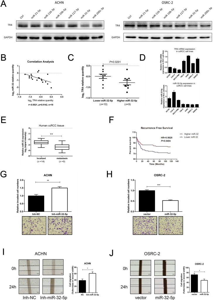
Figure 4. Expression of miR-32-5p’s in ccRCC tissue samples and effects on invasion and migration in vitro.
(A), Some screened miRNAs that could target TR4 are shown using immunoblot analysis in ACHN (left) and OSRC-2 (right) cells. (B), Correlation analysis of miR-32-5p and TR4 mRNA levels using Pearson correlation coefficient in 19 ccRCC samples. (C), The 19 ccRCC samples were divided into 2 groups according to their miR-32-5p expression and ccRCC tissues with lower miR-32-5p expression displayed higher TR4 level, and vice versa. (D), The negative correlation tendency of miR-32-5p (lower panel) and TR4 (upper panel) in ccRCC cell lines. (E), The expression of miR-32-5p in ccRCC tissue samples from patients with distant metastases compared to tissues from metastasis-free patients (data from GEO DataSets website). (F), Recurrence-free survival curves of ccRCC patients analyzed according to the miR-32 expression data from TCGA website, (top curve, Higher miR-32 and bottom curve, Lower miR-32). (G,H), Invasion ability of miR-32-5p inhibitor treated ACHN cells (G), and miR-32-5p overexpressed OSRC-2 cells (H) were compared to their control cells using Matrigel-coated Transwell assays. (I-J), Migration ability of miR-32-5p inhibitor treated ACHN cells(I), and miR-32-5p overexpressed OSRC-2 cells (J) were compared to their control cells using wound healing assays. The percentage of decreased wound widths were compared between the control group and experimental group. Each sample was run in triplicate and in multiple experiments. Data presented as mean±SEM. P <0.05 was considered statistically significant. *P < 0.05, **P < 0.01, and ***P < 0.005.

