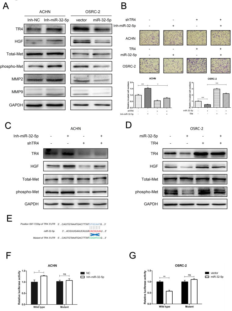
Figure 5. Mechanism dissection how miR-32-5p suppresses ccRCC invasion and migration.
(A), The protein levels of TR4, HGF, Total-Met, phospho-Met, MMP2 and MMP9 were determined by immunoblot analysis after using the miR-32-5p inhibitor in ACHN cells (left) and infection with miR-32-5p in OSRC-2 cells (right). (B), The invasion abilities of ACHN cells (upper panels. +/− shTR4 and +/− miR-32-5p inhibitor) and OSRC-2 cells (middle panels +/− TR4 and +/− miR-32-5p) were determined by Matrigel-coated Transwell assay. Quantitations are on lower panels. (C-D), The protein levels of TR4, HGF, Total-Met and phospho-Met were determined using immunoblot analysis of ACHN cells (c, +/− shTR4 and +/− miR-32-5p inhibitor) and OSRC-2 cells (d, +/− TR4 and +/− mir-32-5p). (E), Sequence alignment of the TR4 3'UTR with potential wild type versus mutant miR-32-5p targeting sites. (F-G), Luciferase reporter activity after transfection of wild type TR4 3’UTR reporter construct in the ACHN (F) and OSRC-2 (G) cells treated with inhibitor of miR-32-5p or overexpressing miR-32-5p, respectively, vs control cells. Data presented as mean±SEM. P<0.05 was considered statistically significant. *P < 0.05, **P < 0.01, ***P < 0.005, ns=not significant.

