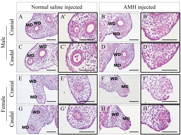
Fig. 7.
HE staining for the urogenital ridges of normal male and normal female in an organ culture (A–H) and magnified images focusing on the Müllerian ducts (A’–H’). The Müllerian ducts with AMH (B, D, F and H) are clearly more regressed than those with normal saline (A, C, E and G) at the same height of the mesonephros, respectively. Scale bars=100 µm.

