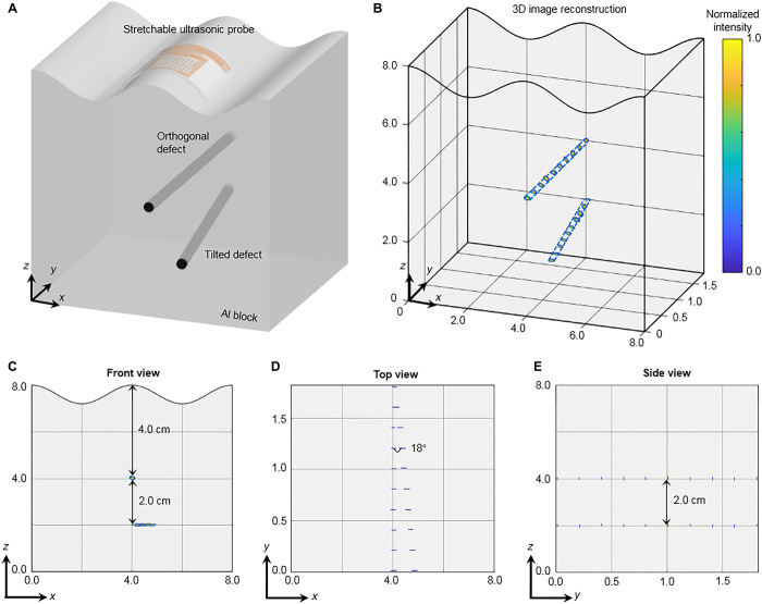
Fig. 5. Three-dimensional image reconstruction of intricate defects under a convex surface.
(A) Schematics of the experimental setup, illustrating the spatial location and relative orientation of the two defects in the test subject. (B) The reconstructed 3D image, showing complete geometries of the two defects. (C to E) The 3D image from different view angles, showing the relative positions and orientations of the two defects to the top surface, which match the design well.

