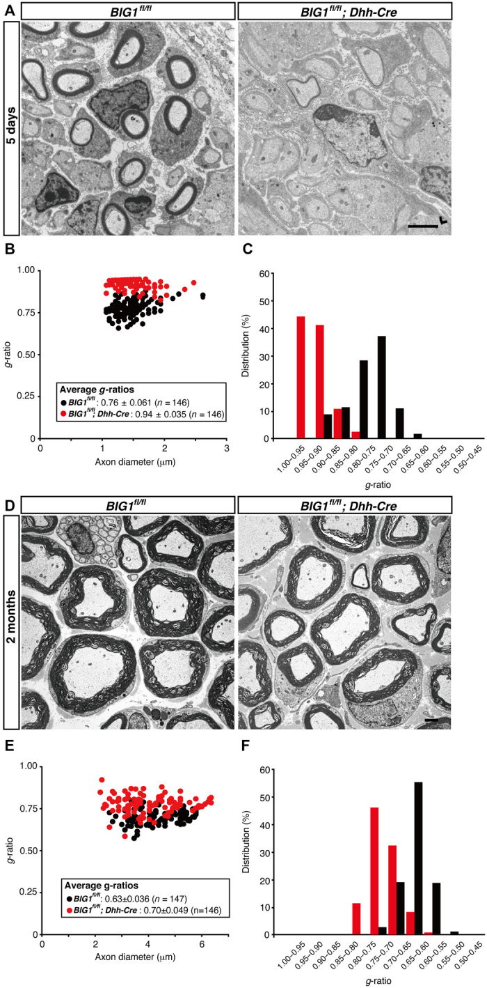
Fig. 2. Dhh-Cre–mediated BIG1 knockout mice exhibit decreased myelin thickness.

(A) Electron microscopic images of sciatic nerve cross sections of conditional knockout (BIG1fl/fl; Dhh-Cre) and control mice at 5 days postnatal are shown. Scale bar, 1 μm. (B and C) A graph of g-ratios of myelinated axons for axon diameters, as well as their distributions, is shown (n = 146 nerves for knockout mice and n = 146 nerves for controls; three independent mice). (D) Electron microscopic images of sciatic nerve cross sections of conditional knockout and control mice at 2 months postnatal are shown. Scale bar, 1 μm. (E and F) A graph of g-ratios of myelinated axons for axon diameter, as well as their distributions, is shown (n = 146 nerves for knockout mice and n = 147 nerves for controls; three independent mice).
