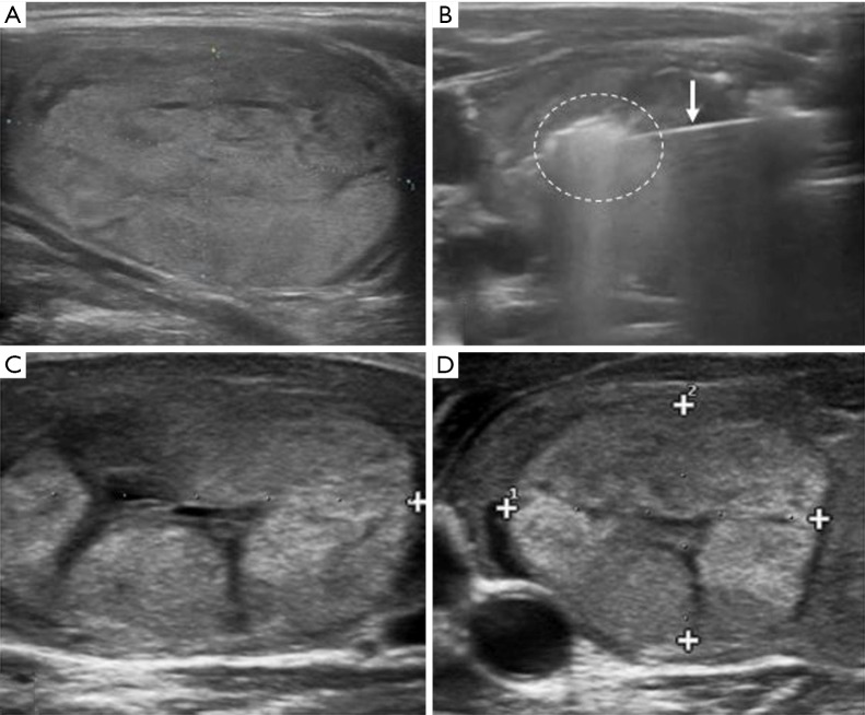
Figure 2.
RFA procedure of a benign thyroid nodule. (A) US image of a thyroid nodule of the right lobe in a 47-year old woman; (B) RFA of the nodule: the needle inside the lesion (white arrow) with the appearance of a hyper echogenic area represented the ablated area; (C) US appearance of thyroid nodule after RFA treatment at 1 month follow-up. Reduction in nodule volume can be appreciated; (D) US image shows a significant shrinkage of the benign nodule compared to image (A) and (C) at 6-month follow-up study. +, the cursors to measure the lesion diameter. US, ultrasound; RFA, radiofrequency ablation.

