
Fig. 3.
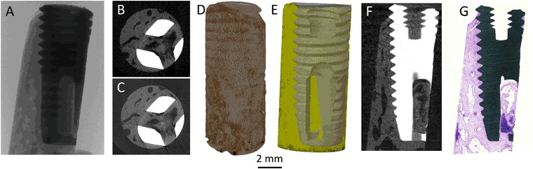
Macro level of osseointegration: Different views of a micro-CT analysis starting with a X-ray imaging. b Typical reconstruction of a low-resolution scan, 12 µm voxel size (typically 15 min). c Typical reconstruction of a high-resolution scan, 2.5 µm voxel size (typically 8 h). d A three-dimensional volume rendering of the reconstructed image stack. e A three-dimensional surface rendering of the segmented data set representing the implant (grey) and bone (yellow) which has been made semi-transparent. f Data set rotated to locate the slice obtained as the histological ground-section. g Overview image of the ground-section stained by toluidine blue