Figure 1.
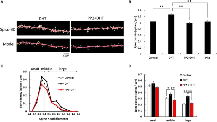
Effects of Src kinase blocker (PP2) on DHT-induced spine increase and change in morphology in hippocampal slices. (A) Spines were analyzed along the secondary dendrites of pyramidal neurons in the stratum radiatum of CA1 neurons. Dendrite after DHT-treatment for 2 h (DHT) and dendrite after DHT plus PP2 treatment for 2 h (PP2+DHT). (Spiso) shows the image of dendrite and spines analyzed with Spiso-3D software. Maximal intensity projections onto XY plane is shown. Traced dendrite is shown in red color and spines are indicated in yellow color. (Model) shows 3 dimensional model illustration of (Spiso) image. Bar, 5 μm. (B) Effect of treatments by DHT or PP2 on the total spine density in CA1 neurons. Vertical axis is the average number of spines per 1 μm of dendrite. A 2 h treatment in ACSF without drugs (Control), with 10 nM DHT (DHT), with 10 nM DHT and 10 μM PP2 (PP2 + DHT), and with PP2 only (PP2). (C) Histogram of spine head diameters after a 2 h treatment in ACSF without drugs (Control, black dashed line), with 10 nM DHT (black line), with 10 nM DHT and 10 μM PP2 (red line). Spines were classified into three categories depending on their head diameter, e.g., 0.2–0.4 μm as small-head spines, 0.4–0.5 μm as middle-head spines, and larger than 0.5 μm as large-head spines. Vertical axis is the number of spines per 1 μm of dendrite. (D) Density of three subtypes of spines. Abbreviations are same as in (B). Vertical axis is the number of spines per 1 μm of dendrite. From left to right, small-head spines (small), middle-head spines (middle), and large-head spines (large) type. ACSF without drugs (Control, open column), DHT (black column), PP2 + DHT (red column). Vertical axis is the number of spines per 1 μm of dendrite. Results are represented as mean ± SEM. Statistical significance yielded *P < 0.05, **P < 0.01 vs. DHT sample. For DHT, PP2+DHT, and PP2 only treatments, we investigated, 50 dendrites with 2,300–2,700 spines from 3 rats, 12 slices, and 30 neurons. For control, we used 80 dendrites with ~4,000 spines from 6 rats, 24 slices and 50 neurons.

