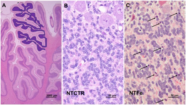
Figure 1.

Photomicrography of histological features. (A) Assessed area of the internal granular cell layer of three complete gyri of the anterior cerebellar lobe as highlighted in blue. (B,C) Representative images of the inner granular cell layer of the NTCTR and NTFe groups are shown. Arrows indicate cells with homogenous eosinophilic cytoplasm and pyknotic nuclei.
