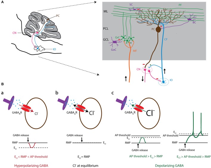
Figure 1.
GABAergic signaling in olivo-cerebellar circuit. (A) Schematic representation of a sagittal section of the mouse olivo-cerebellar system (left). Inferior olivary neurons (shown in blue) project to the cerebellar cortex (gray) and excite Purkinje cells (brown), as well as the deeply located cerebellar nuclei (CN) neurons (pink). A particular subset of CN neurons projects back to the inferior olive (IO), forming the olivo-cortico-nuclear loop. The right panel demonstrates the anatomical circuit of the cerebellar cortical neurons and their connectivity with CN and the IO. ML, molecular layer; PCL, Purkinje cell layer; GCL, granule cell layer; PC, Purkinje cell; GrC, granule cell; SC, stellate cell; BC, basket cell; GoC, Golgi cell; PF, parallel fiber; CF, climbing fiber; MF, mossy fiber; CN, cerebellar nuclei; IO, inferior olive. (B) The level of intracellular chloride concentration ([Cl−]i) dictates the polarity of the current through GABAA receptors (GABAARs). If [Cl−]i is low, the reversal potential of Cl− (ECl) becomes negative compared to the resting membrane potential (RMP). In this condition GABAARs mediate an inward Cl− current that results in hyperpolarization of the cell membrane (a). In contrast, high [Cl−]i results in a positive shift of ECl and leads to an outward Cl− current through GABAAR and depolarization of the cell membrane that potentially induces action potential firing (c). In conditions where ECl shifts to values similar to RMP, there will be no net Cl− current through GABAARs (b).

