
Figure 4.
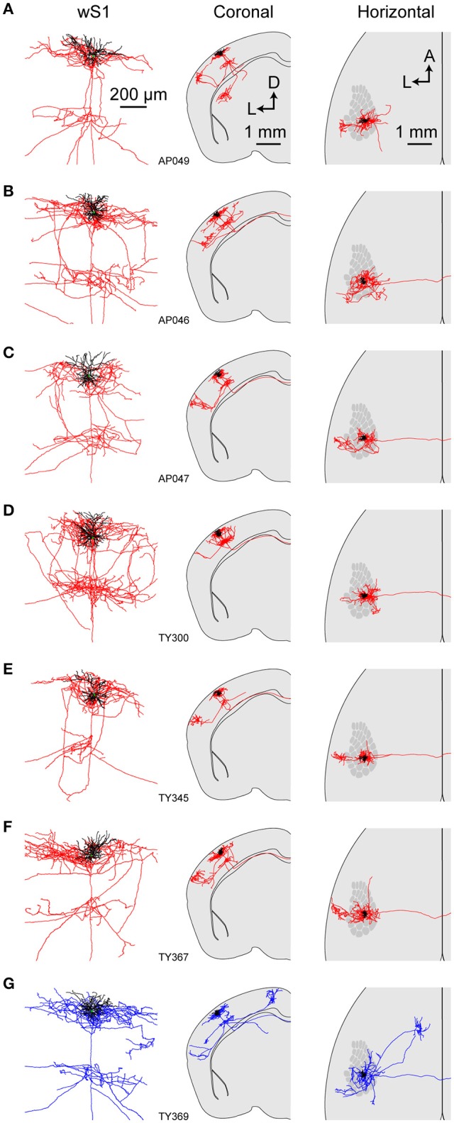
Axonal and dendritic structure of neurons retrogradely-labeled from wS2. (A–G) Dendritic (black) and axonal (red in A–F, blue in G) arborisations of different individual neurons viewed locally in wS1 (left), in coronal projection (center) and in horizontal projection (right). The wS1 barrel field is schematically indicated (right).