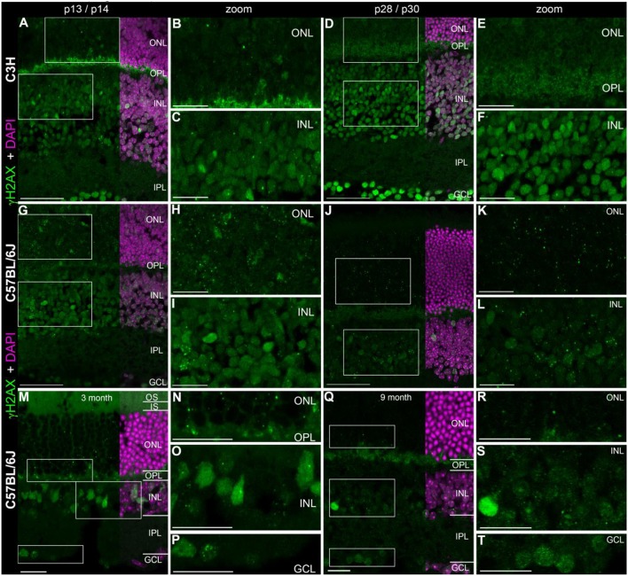
Figure 1.
Presence of γH2AX in wildtype retina at different ages. (A) Entire retina and magnifications of ONL (B) and INL (C) of C3H mice at 2 weeks of age. (D) Entire retina and magnifications of ONL (E) and INL (F) of C3H mice at 4 weeks of age. (G) Entire retina and magnifications of ONL (H) and INL (I) of C57BL/6J mice at 2 weeks of age. (J) Entire retina and magnifications of ONL (K) and INL (L) of C57BL/6J mice at 4 weeks of age. (M) Entire retina and magnifications of ONL (N), INL (O), and GCL (P) of C57BL/6J mice at 3 months of age. (Q) Entire retina and magnifications of ONL (R), INL (S), and GCL (T) of C57BL/6J mice at 9 months of age. For detailed description see Results. IS, inner segments; ONL, outer nuclear layer; OPL, outer plexiform layer; INL, inner nuclear layer; IPL, inner plexiform layer; GCL, ganglion cell layer. Scales A,D,G,J: 50 μm, scales B,C,E,F,H,I,K,L,M–T: 20 μm.

