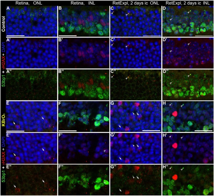
Figure 8.
Co-localization of γH2AX and 53bp1 following double strand break induction with KBrO3. γH2AX and 53BP1 double labeled foci were found only in retinal explant culture, in both the KBrO3 treated and untreated preparations (the two right pannels, arrow heads). Nuclei with several γH2AX immunoreactive foci were found in the ONL of KBrO3 treated tissue only (arrows). DAPI counterstaining (blue) reveals the chromatin in relation to the location of γH2AX and 53BP1 immunoreactive foci in the nuclei. (A–B”) Vertical frozen sections of control retina of 3-month-old mice. (E–F”) Sections of 3-month-old mice after KBrO3 incubation. (C–D”) Sections of control retina of retinal explants after 2 days in culture. (G–H”) Sections of retinal explants treated with KBrO3 after 2 days in culture. ONL, outer nuclear layer; INL, inner nuclear layer. All scales = 20 μm.

