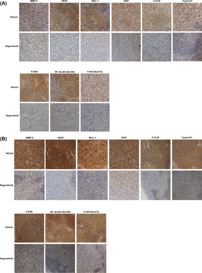
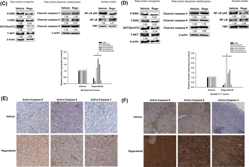
Figure 3. Effect of regorafenib on expression of P-ERK, AKT (Ser473), NF-κB p65 (Ser473), and NF-κB-modulated downstream effector proteins in SK-Hep1/luc2 tumor and Hep3B 2.1-7 bearing mice.
Mice were killed on day 14 after treatments and protein levels in tumor tissues were evaluated with IHC staining. (A) Protein levels of MMP-9, VEGF, MCL-1, XIAP, C-FLIP, Cyclin-D1, and P-ERK, AKT (Ser473), NF-κB p65 (Ser473) on SK-Hep1/luc2 tumor by IHC. (B) IHC staining of Hep3B 2.1-7 tumor. (C) Phosphorylation oncogenes and apoptosis-related cleavage proteins expression which validated by Western blotting on SK-Hep1/luc2 tumor. (D) Western blotting of Hep3B 2.1-7 tumor. (E) Expression of antiapoptotic proteins (active Capase-9, -8, and -3) on SK-Hep1/luc2 tumor by IHC. (F) IHC staining of Hep3B 2.1-7 tumor; aP<0.01 as compared with vehicle group; C-FLIP, cellular FADD-like IL-1β-converting enzyme-inhibitory protein; IHC, immunohistochemistry; MCL, myeloid leukemia cell differentiation protein; MMP, matrix metallopeptidase; NF-κB, nuclear factor-κB; P-ERK, phosphorylated extracellular signal-regulated kinase; VEGF, vascular endothelial growth factor; XIAP, X-linked inhibitor of apoptosis protein.


