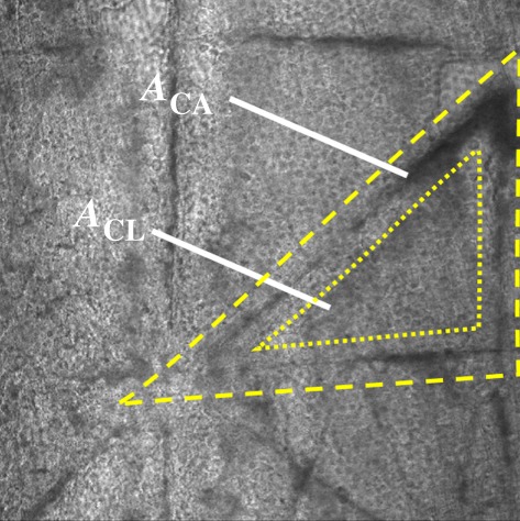
Figure 3.

Characterizing regions of SC tissue. Transmitted light image of isolated human SC showing triangular subunits. The area in between the dotted and dashed line corresponds to the topographical canyon region, ACA, while the region within the dotted line corresponds to the inner cluster region, ACL. (Online version in colour.)
