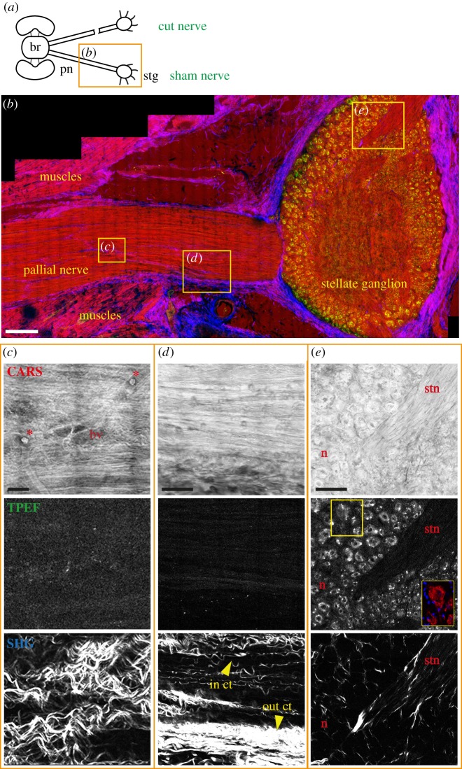
Figure 1.
Sham control nerve. (a) Schematic drawing showing the connections between the brain (br) and stellate ganglion (stg) through the right and left pallial nerves (pn). The orange rectangle delimits the area of the nerve shown in the figure. The sham nerve is shown in (b) after multiphoton imaging. CARS signals (red in b) highlighted the structure of axons in the pallial nerve (b–d) and the neuropil, neurons (n) and stellar nerves (stn) inside the stellate ganglion (b,e). CARS signal also showed blood vessels (bv) and haemocytes (red asterisks) in the nerve (c). In (b) also muscles appear to give a strong signal in CARS. SHG (blue in b) allowed visualization of the outer connective tissue (out ct) enwrapping the pallial nerve (b,d) and the stellate ganglion (b), and the inner connective tissue (in ct) surrounding bundles of fibres inside the pallial nerve (c,d). TPEF signal (in green) was strongly detected only in the neurons of the stellate ganglion (e). Neurons were identified via acetylated tubulin labelling, visible in red in the yellow box in (e). Glial cells around neurons are not highlighted using multiphoton microscopy, but their nuclei are detected via DAPI counterstain (blue in the yellow box in e). Scale bars: (b) 250 µm, (c) 20 µm, (d,e) 50 µm.

