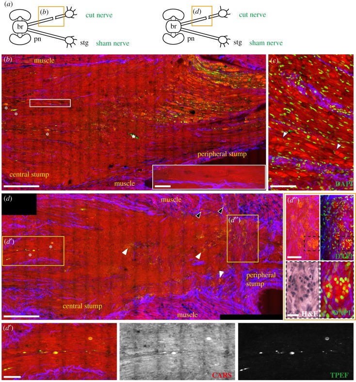
Figure 6.
Injured nerve 14 days post lesion. (a) Schematic drawing showing the connections between the brain (br) and stellate ganglion (stg) through the right and left pallial nerves (pn). The orange rectangle delimits the area of the nerve shown in the figure panel. (b,d) The central and peripheral stumps of a nerve 14 days post lesion. The majority of the fibres in the peripheral stump appear swollen and fragmented, while the few degenerating axons in the central stump are marked with white asterisks (also visible in d′, CARS and TPEF). Small granules, arranged in rows are visible in the central stump (enlargement in the white rectangle in b). In (c) the peripheral stump is counterstained with DAPI, showing that cell nuclei are outside the ovoidal fragments and not infiltrating them. Some of these fragments contain a structure resembling a nucleus (marked with white arrows) which, however, does not stain with DAPI. Drop shaped structures were identified in the lesioned nerve (white arrows in d; enlargement in d″) and muscles (black arrows in d), highlighted by CARS and TPEF (red and green in d). Counterstain with DAPI allowed identification of those structures as cell clusters (d″) surrounded by connective tissue (d″ SHG). H&E staining (black dotted box in d″) slightly stained these structures. White stars in figure (b) indicate artefacts. Scale bars: (b,d) 250 µm, (c) 100 µm, (d′,d″) 50 µm, (e) 20 µm, white rectangle in (b) 25 µm.

