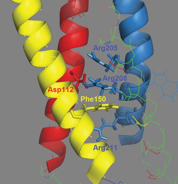
Figure 3.

Side view of the open human HV1 channel, with the external end up. Transmembrane helices are colour-coded: S1 = red, S2 = yellow, S4 = blue and S3 is shown as lines to be unobtrusive. Key amino acids are labelled and shown with side chains as sticks. Asp112 is crucial for selectivity; Phe150 demarcates inner and outer aqueous vestibules and the three Arg in S4 sense voltage. Figure is based on the model of Li et al. [91]. Note that Asp112 interacts with Asp208 [94], and Arg211 is below Phe150, and thus is exposed to the inner vestibule. Drawn with PyMol.
