
Figure 1. Identification of a truncating mutation in PATL2 .
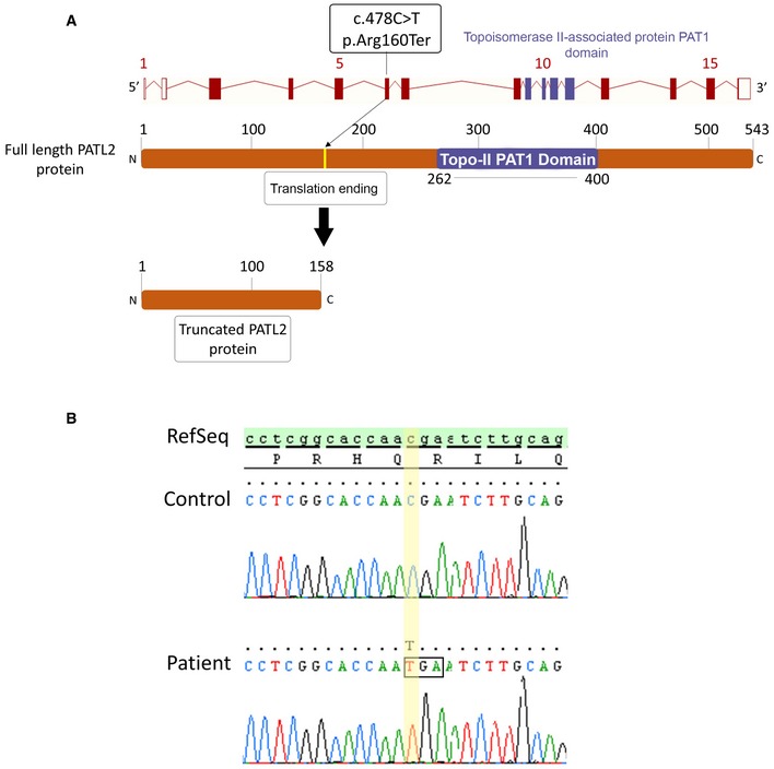
- Location of the PATL2 mutation in the intron–exon structure and in a representation of the corresponding amino acid sequence. The variant identified, homozygous in the six patients, is located in exon 6 and creates a STOP codon, ending translation and producing a truncated 158‐amino acid (aa) protein instead of the full‐length 543 aa, and lacking the essential PAT1 (topoisomerase II‐associated protein PAT1) domain.
- Electropherograms of Sanger sequencing for patients harbouring PATL2 mutations compared to reference sequence.