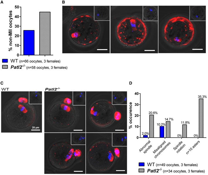
Figure 5. Infertility of Patl2‐deficient female mice is due to oocyte maturation defects.

- Oocytes collected after ovarian stimulation were labelled with a tubulin antibody (red) and counterstained with DAPI to reveal DNA (blue). An increase in non‐MII oocytes (MI arrest, absence of PB) after full ovarian stimulation was observed in Patl2 −/− mice (n = 3 females per genotype).
- IF images of tubulin‐stained Patl2 −/− oocytes arrested at MI stage showing various defects such as irregular spindle shape and abnormal chromosome distribution. Scale bar = 20 μm. Inset in each panel shows overlay of phase contrast image and Hoechst staining of the corresponding oocyte. No polar bodies were observed for MI oocytes.
- IF images of tubulin‐stained WT and Patl2 −/− MII oocytes, as evidenced by PB1. In control MII oocytes, stack projections of confocal images show that the spindle was symmetric and the chromosomes distributed in the middle of the spindle. In contrast, in Patl2 −/− MII oocytes various defects were observed such as irregular spindle shape, spindle rotation and numerous cytoplasmic asters. Slightly greater numbers of oocytes with abnormal chromosome distribution were also observed. Scale bar = 20 μm. Inset in each panel shows overlay of phase contrast image and Hoechst staining of the corresponding oocyte. One polar bodies was observed for MII oocytes.
- Histograms quantifying the % defects observed in Patl2 −/− MII oocytes.
