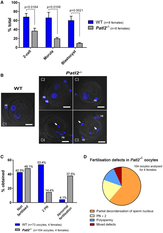
Figure 6. MII oocytes from Patl2 −/− female mice exhibit abnormal fertilisation preventing normal embryo development.

-
AIVF outcomes (mean ± SEM) measured at the two‐cell, morula and blastocyst stages show that the developmental competence of Patl2 −/− oocytes is compromised. Oocytes were collected from stimulated WT and Patl2 −/− females and sperm from WT males. For each IVF replicate (different WT males), IVF outcomes at different stages were compared for WT and Patl2 −/− oocytes. Statistical differences were assessed using unpaired two‐tailed t‐tests.
-
BZ‐stack projections of confocal images of 2PN zygotes obtained from WT and Patl2 −/− oocytes 6‐8 h after sperm–egg mixing. Fertilised WT oocytes exhibit normal 2PN stage (C1) whereas the number of fertilised Patl2 −/− oocytes exhibiting normal 2PN stage is strongly reduced (C2) and most of them show defects such as absence of a male pronucleus (C3), partial decondensation of male PN (C4, C5, white arrows) or polyspermy (C5 white arrows). Scale bar = 20 μm, PB = polar body, PN = pronucleus.
-
C, DThe percentage of 2PN obtained 6–8 h after fertilisation drops from 53.4% for WT to 14.4% for Patl2 −/− oocytes, which exhibit various fertilisation abnormalities including partial decondensation of sperm DNA, polyspermy, abnormal number of PN or mixed defects (D).
