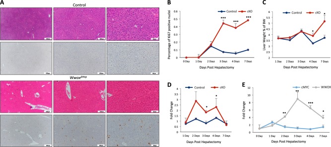
Fig. 7. Wwox ablation is associated with increased proliferation upon partial hepatectomy.
a Histological images (H&E staining and Ki67 immunohistochemical staining) of control and WwoxΔHep mice liver 3-days post 50% partial hepatectomy (× 10, × 20, and × 40). b Quantification of positive Ki67 nuclei in 5 months old control (WT) and WwoxΔHep mice liver 1, 2, 3, 4, and 7 days post hepatectomy (n = 5 for each group). c Liver weight to body weight ratio of 1, 2, 3, 4, and 7 days post hepatectomy (n = 5 for each group). d mRNA expression levels of c-Myc in control and WwoxΔHep mice liver 1, 2, 3, 4, and 7 days post hepatectomy. e mRNA expression levels of Wwox in control and WwoxΔHep mice liver 1, 2, 3, 4, and 7 days post hepatectomy (n = 4 for each group). * P value < 0.05, ** P value < 0.01, *** P value < 0.001. Error bars indicate ± SEM

