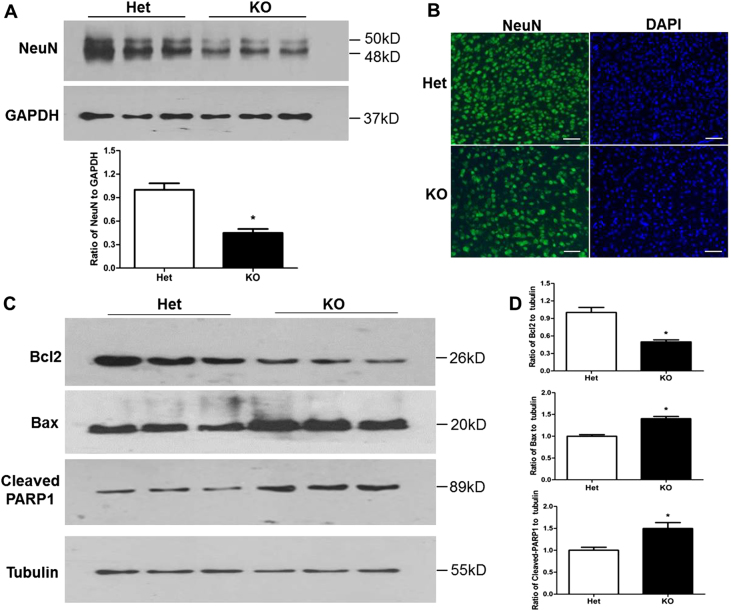
Fig. 4. Tctn3 KO led to altered apoptosis-related protein levels in mouse brains.
At E12.5, mouse brains were collected, and protein levels of NeuN were determined in Tctn3 Het KO mice and Tctn3 KO mice by Western blot analysis (a). At E12.5, mouse brains were fixed, and immunofluorescent staining was performed using anti-NeuN antibody (b). The levels of apoptosis-related proteins Bcl2, Bax, and cleaved PARP-1 were examined by Western blot analysis. Beta-tubulin was used as a loading control. *P < 0.05 vs Het mice. N = 6. Scale bars: 10 μm

