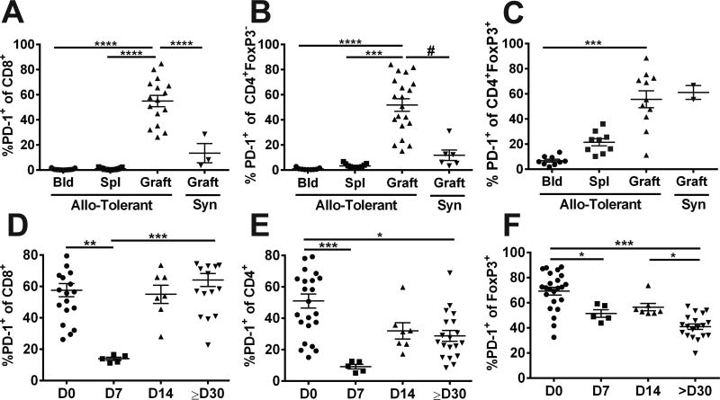
Figure 5.
Listeria infection of tolerant mice results in transient reduction in the percentages of PD1-expressing CD8+ and CD4+Foxp3− cells in the allograft. (A-C) Percentage PD-1+ of CD8+, CD4+Foxp3− and CD4+Foxp3+ cells in the blood, spleen and infiltrating BALB/c allografts in tolerant C57BL/6 mice, or syngeneic grafts at ≥ 60 days post-transplantation. (D-F) Percentages of PD-1+ of CD8+CD4+Foxp3+CD4+Foxp3− and CD4+Foxp3+ cells in the tolerant allografts, prior to (D0) and 7, 14 and ≥30 days post-Listeria infection. Each symbol represents a single animal, data are presented as mean ± standard error, and statistical significance at p≤ *0.05, **0.01, ***0.001 and ****0.0005 by Kruskal-Wallis multiple comparison test, and p≤#0.05 by pair-comparison. Detailed gating strategy for PD-1 expression by each of the T cell subset is illustrated in Figure S2.

