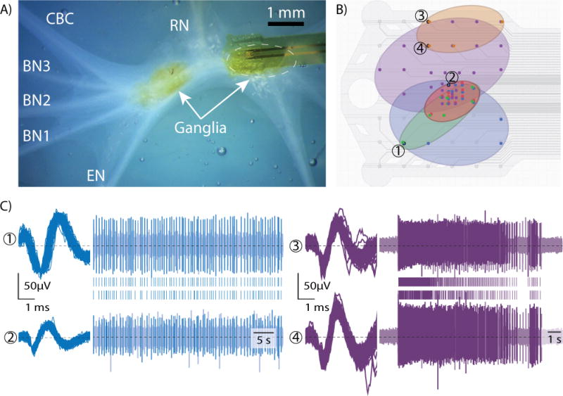
Figure 2.
A) Brightfield microscope image of array on buccal ganglia, with radular nerve (RN), buccal nerves (BN1-3), cerebral-buccal connective nerve (CBC), esophageal nerve (EN) and ganglia labeled (right ganglion obscured by array) B) Array schematic with 5 regions of similarly firing channel overlaid C) Sample waveforms, raw voltage traces, and raster plots [numbers and colors correspond to those in (B)] Note that the right (purple) unit is on channels with a second unit also recorded, which corresponded to the orange region

