Figure 10.

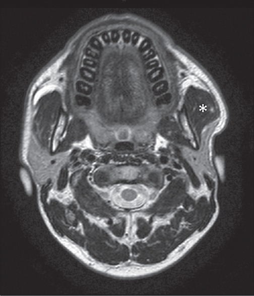
A 37-year-old female patient with swelling of the left preauricular region, negative at ultrasonography scan. Axial T2-weighted magnetic resonance scan easily demonstrates a unilateral hypertrophy of the left masseter muscle (asterisk).

A 37-year-old female patient with swelling of the left preauricular region, negative at ultrasonography scan. Axial T2-weighted magnetic resonance scan easily demonstrates a unilateral hypertrophy of the left masseter muscle (asterisk).