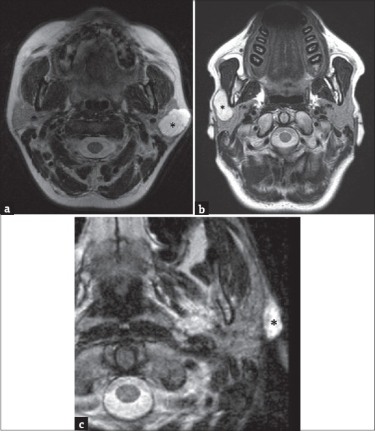
Figure 2.

(a) A 53-year-old female axial T2-weighted magnetic resonance image showing the presence of a high-signal-intensity lesion (asterisk) in the left parotid gland. This lesion arises from the superficial lobe, presenting a partial exophytic development. (b) A 44--old male axial T2-weighted magnetic resonance scan in another patient showing a well-defined hyperintense lesion arising in the right superficial parotid lobe (asterisk); the lesion occupies the anterior extension of parotid gland superficially to the masseter muscle. In such cases, the only ultrasonography examination was not accurate enough to define the origin of lesions, while cross-sectional imaging identifies the exact location and their adjacent and distant involvement. (c) A 62-year-old male – T2-weighted axial magnetic resonance image shows an extraglandular lesion (asterisk), clearly originating from the nearby tissues.
