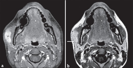
Figure 7.

(a) A 66-year-old male axial T1-weighted fat-suppressed magnetic resonance image obtained after intravenous gadolinium administration. The images show the contrast enhancement of a lesion (asterisk) arising from the masseter muscle. (b) A 66-year-old male axial T1-weighted fat-suppressed magnetic resonance image obtained after intravenous gadolinium administration. The images show the contrast enhancement of a lesion (asterisk) arising from the masseter muscle.
