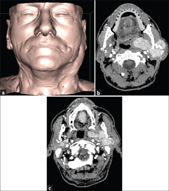
Figure 8.

(a) A 76-year-old male three-dimensional-shaded surface display reformatted image of a patient with swelling of the left preauricular region. (b) A 76-year-old male axial contrast-enhanced computed tomography scans showing the presence of several lesions of the left parotid gland, detectable from the superficial lobe to the adjacencies of the parapharyngeal space. (c) A 76-year-old male concurrent lesions in the deep lobe of the right parotid gland can also be seen. In this patient, a previous US undervalued the number of the lesions, probably because of their deep location (i.e., deep lobe of the parotid gland or behind the shadow of the mandible).
